Learning is
an act of curiosity.
- Aorta: Main artery
- Pulmonary trunk: Carries used blood to the lungs
- Superior vena cava: Brings blood back from head and arms
- Inferior vena cava: Brings blood back from everything below
- Pulmonary veins: The only veins carrying oxygen-rich blood
- Left Atrium: Receives oxygenated blood
- Right Atrium: Receives venous blood
- Right auricle: Ear-shaped pouch on the right atrium
- Left auricle: Where clots form in atrial fibrillation
- Left Ventricle: Pumps to the body
- Right Ventricle: Pumps to the lungs
- Apex: The tip you feel beating on the chest
- Anterior interventricular groove: Traces the wall between the ventricles
- Coronary groove: The ring dividing atria from ventricles
- Left coronary artery: Feeds most of the left ventricle
- Right coronary artery: Feeds the right side and the pacemaker
- Anterior interventricular branch: Block it and the front wall dies
- Circumflex branch: Curves round to the back of the left side
- Right marginal branch: Runs along the lower right border
- Great cardiac vein: Runs beside the front artery, going back
- Coronary sinus: One collector empties it all into the right atrium
- Aortic arch: Turns the aorta backwards and down
- Brachiocephalic trunk: First branch: right arm, right side of head
- Left common carotid artery: Second branch: the left side of the head
- Left subclavian artery: Third branch: the left arm
- Descending aorta: Carries on down behind the heart
- Right pulmonary artery: To the right lung
- Left pulmonary artery: To the left lung
- Mitral Valve: Prevents backflow
- Tricuspid valve: Between right atrium and right ventricle
- Aortic valve: Stops blood falling back from the aorta
- Interventricular septum: The muscular wall between the two pumps
3D specimen · click a dot to exploreCor
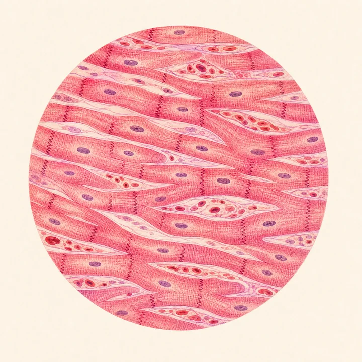
Microscopic view
Cardiac muscle tissue
Compare organs
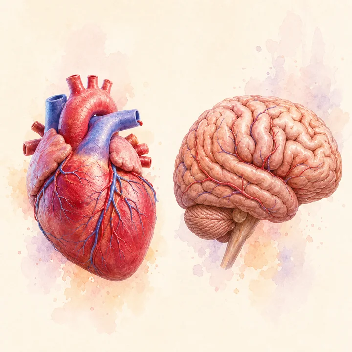
Heart vs. brain
Function animation
Circulates oxygenated blood
Clinical notes
Common conditions
- MyocarditisDiffuse
I40.9 - Congenital heart defectsDiffuse
Q24.9
Where it works